№ 01
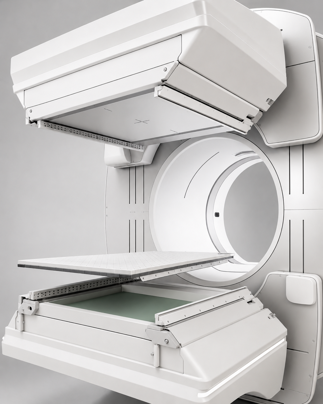
A dual-head clinical SPECT platform — full collimator set.
Low-, medium-, and high-energy classes. Parallel-hole and pinhole geometries. Continuous program supply since 1994.
32 yrs
Active
Active
Collimators, mechanical assemblies, machined components, and accessory equipment for nuclear medicine imaging systems. Engineering consultation, global sourcing, and hard-to-find spare parts for legacy and active platforms. A Tier 1 manufacturing partner to the world's leading SPECT and gamma-camera OEMs — since 1993.

A SPECT scanner forms its image from gamma photons emitted inside the patient. Those photons travel in every direction at once — an unfiltered detector would record only noise.
The collimator is the precision-machined lead lens that solves this. A honeycomb of microscopic channels mounted on the inner face of each detector head, admitting only the photons travelling on a true perpendicular path. It is the component that defines spatial resolution, sensitivity, and image geometry — the physical limit of what the system can see.
Channel pitch and septa thickness set the smallest detail the system can resolve.
Hole geometry governs how many true photons reach the crystal per unit time.
Parallel, fan-beam, or pinhole optics shape how the patient is projected onto the detector.
A small number of programs, served at depth. Direct Tier 1 supply to the world's leading SPECT and gamma-camera platforms — on systems shipping today and on installations still in clinical service twenty years on.
Low-, medium-, and high-energy collimators across every standard geometry — engineered to the customer's drawing, focal geometry, and detector format. Each unit inspected against the program's own acceptance criteria.

Identical channels perpendicular to the detector. Only true-normal photons survive the septa; the image is geometrically faithful at 1:1.
Whole-body bone, lung perfusion, renal, brain SPECT.
The standard general-purpose geometry. Low, medium, and high energy across the diagnostic isotope range.

Channels converge toward a focal line; the projection magnifies in one axis while preserving 1:1 in the other — trading field of view for detail.
Myocardial perfusion SPECT, brain SPECT (DaTscan, perfusion).
Converging-geometry optics for magnified cardiac and small-organ acquisitions. To customer focal length.

Photons cross at a single aperture and form an inverted image on the detector. Magnification is geometric — set by focal and object distance.
Thyroid, parathyroid, sentinel node, paediatric and small-animal SPECT.
Single-aperture optics for the highest spatial resolution on small targets.

Rectangular-section channels perpendicular to the detector. The long axis raises geometric efficiency for slot-shaped targets; the short axis preserves resolution.
Whole-body planar studies, lymphoscintigraphy, high-throughput scans.
Slot-channel geometries for high-sensitivity acquisitions. To customer pitch and interface.
Precision extrusion, machining of solid lead castings, bonding, and complex mechanical assembly — the full production chain under one roof, every unit traced to its lot.
Lead castings and structural components, sourced and qualified to specification.
Frame, matrix, and interface assembled to the customer's drawing.
Every unit measured against the program's inspection plan. Documented, lot-traceable, released.
Manufacturing and engineering distributed across dedicated sites, with qualified production partners on three continents. A single program, served end to end.

Share your specification — we will engage as a manufacturing partner from first review through serial production.